Where Is the Brain’s Reward System and How Does It Work?
We report often here at AddictionNews about the brain’s reward system. Today we’ll take a step back and see what researchers are saying about where the brain’s reward system is and how it functions.
A study in the American Journal of Psychiatry attempted to measure what happens in the brains of adolescents when their reward systems are stimulated and how that might relate to the likelihood of developing depression at a later date. The authors begin by claiming the relationship between reward system dysfunction and depression is “well-known”:
Reward system dysfunction is a well-known correlate and predictor of depression in adults and adolescents, with depressed individuals showing blunted (hyporeactive) striatal response to monetary rewards.
There is a long history of using monetary rewards to map the brain’s reward system, from the anticipation of a reward to experiencing the reward and finally the aftermath of the reward. The effects are measured through fMRI brain scans that indicate which areas of the brain “light up” in response to anticipation and consumption of a reward.
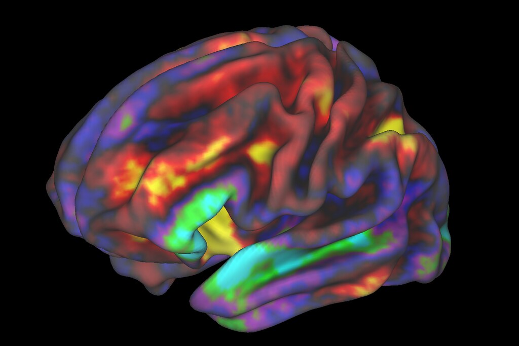
A meta-analysis of scientific papers involving monetary rewards and fMRI brain scans found that 45 of the studies showed reward-based activation in “the ventral striatum, the middle cingulate cortex/supplementary motor area and the insula” while 28 studies showed “ventral striatal activation, plus cortical activations in the anterior and posterior cingulate cortex.” Here’s a link to a colorful map showing which areas of the brain light up when a reward is delivered.
In the American Journal of Psychiatry study involving fMRI on 131 adolescents, “Current depression severity was associated with hyporeactivity exclusively in the nucleus accumbens in response to the anticipation of a reward, while cumulative depression severity was associated with blunted response to anticipation across a cortico-striatal circuit (striatum, ACC, insula).”
The scans were performed on people who had been annually assessed for psychiatric symptoms since early childhood. The scans reveal that “recent depression severity during adolescence was associated with more focal hyporeactivity in the nucleus accumbens, while depression severity during early childhood (i.e., preschool) was associated with more global hyporeactivity across the cortico-striatal circuit.”
The way the rewards are administered and received is interesting. Researchers use a “card guessing game” in which they were able to indicate the person is likely to win or likely to lose, or neither win nor lose, or neutral. This allowed them to measure anticipation. Then they would discover if they won, lost, or no outcome, so they could measure the reward.
The results helped the researchers tease out the difference in the brain’s reward system in response to current depression vs. cumulative depression. Not surprisingly, they found cumulative depression activates a broader neural pattern and current depression was more narrowly focused on “activation in the nucleus accumbens.”
The authors speculate that the study reveals the possibility of a “scar of chronic depression” that begins with a focus in the nucleus accumbens and spreads, over time, so that the “repeated experience of depression that starts early in childhood could lead to downstream hyporeactivity of the entire circuit.” This, in turn, leads to the conclusion that when depression becomes a “habit” it blunts the reward response.
We’ll continue looking at the brain’s reward response system this week at AddictionNews. Stay tuned!
Written by Steve O’Keefe. First published April 16, 2024.
Sources:
“Brain Reward System Dysfunction in Adolescence: Current, Cumulative, and Developmental Periods of Depression,” American Journal of Psychiatry, April 2020.
“Brain activations associated with anticipation and delivery of monetary reward: A systematic review and meta-analysis of fMRI studies,” Plus One, August 5, 2021.
Image Copyright: NIH Image Gallery, used under Creative Commons 2.0 license.